
Machine learning tools and experiments at the ALS enabled the identification of defect-rich regions in single-crystalline Co3Sn2S2 that link to how surface electrons move. Atom-level understanding of how the surface electronic properties of a magnetic semimetal can be tuned could guide its use in advanced technologies like spintronics and catalysis. Read more »![]()
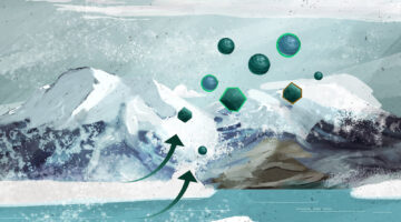
Aerosol Chemistry Offers Clues to the Arctic’s Future
Researchers used scanning transmission x-ray microscopy to analyze Arctic aerosols, which strongly influence cloud formation and overall climate. Understanding what these particles are and how they change as they travel could help improve climate models and yield more accurate predictions of the changing Arctic environment’s global impact. Read more »

February 2026 Message from the UEC Chair
Welcome from the 2026 ALS Users’ Executive Committee (UEC)! As the group dedicated to representing the voice of the ALS user community, our primary role is to bridge the gap between your scientific needs and the operational realities of ALS management. Read more for the outlook on the year and be sure to suggest user meeting speakers and workshops. Read more »
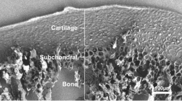
How Zinc Alters Mineral Structure in Early Arthritis
Using high-resolution x-ray techniques, researchers from UCSF, the ALS, and SSRL uncovered structural evidence that zinc subtly alters bone mineral in vulnerable joint regions, revealing early changes that may explain how arthritis begins and progresses. Read more »

2026 Bay Area Light Sources Joint Users’ Meeting
The ALS will join forces with SSRL and LCLS for a Joint Users’ Meeting at SLAC National Accelerator Laboratory, September 20–25. We invite the community to shape the program by nominating plenary speakers and proposing tutorials and workshops. We especially encourage ideas for joint workshops involving ALS, LCLS, and SSRL—whether spanning all three facilities or a collaboration between two. The deadline for proposals is March 13. Read more »