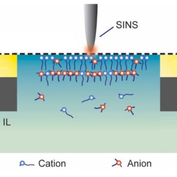
Researchers used infrared spectroscopy at the ALS to detect the molecular behaviors of ionic liquids—which serve as high performance electrolytes in energy storage devices—under varying charge bias conditions. Their insights define a direction for targeted design of ionic liquid-based electrolytes with optimized properties for energy storage applications. Read more »
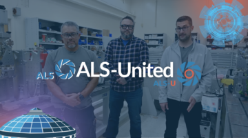
ALS-United: Thomas Gaucher, Raul Mascote, and Adrian Williams
ALS-United is an opportunity to meet the people collaborating at the Advanced Light Source and the ALS Upgrade Project. Hear firsthand how team science enables the cutting-edge research of today and builds the facility of the future. This month, we spoke with Thomas Gaucher (Manufacturing Engineer), Raul Mascombe (Engineering Technical Associate), and Adrian Williams (Engineering Technical Associate). Read more »

Accelerate UX Workshop Brings Global Expertise Together at the ALS
Staff from eleven different accelerator lab facilities gathered at the ALS to improve the user experience for operators, researchers, engineers, and more. Workshop participants learned from experts in the UX field and even took part in a hackathon that paired beamline scientists with interface developers. Read more »
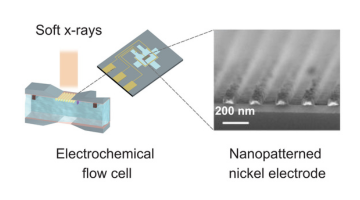
Strength in Numbers: Nanopatterns Amplify X-Ray Signals from Buried Interfaces
Berkeley Lab researchers developed a new x-ray technique that uses nanoscale patterns to amplify weak signals, allowing scientists to observe chemical reactions at buried solid–liquid interfaces that were previously challenging to study. Read more »

Aerosol Chemistry Offers Clues to the Arctic’s Future
Researchers used scanning transmission x-ray microscopy to analyze Arctic aerosols, which strongly influence cloud formation and overall climate. Understanding what these particles are and how they change as they travel could help improve climate models and yield more accurate predictions of the changing Arctic environment’s global impact. Read more »